
Understanding Jaw and Facial Surgery Through Educational Content

Jaw and facial surgery can feel complex before a patient has a full consultation. Terms like orthognathic surgery, Le Fort I, BSSO, double jaw surgery, and genioplasty may appear in videos or before-and-after cases, but they are not always easy to understand without context.
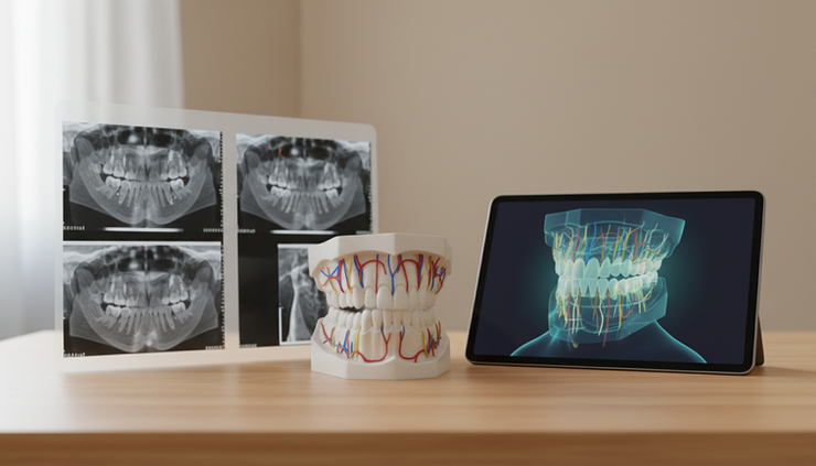
Reliable educational content can make these ideas clearer. Well-written articles, educational videos, 3D simulations, and carefully explained patient cases can help people learn the basic principles of jaw and facial surgery, treatment planning, and recovery. They can also help patients ask better questions during consultation.
At the same time, educational content has limits. It can explain general concepts, but it cannot replace an individualized clinical examination, diagnosis, imaging review, and surgeon-led treatment plan.
What orthognathic surgery means
Orthognathic surgery is corrective jaw surgery that changes the position of the upper jaw, lower jaw, or both. It is usually considered when the jaws do not align properly because of skeletal growth patterns, facial imbalance, bite problems, airway-related concerns, or functional issues that cannot be corrected with orthodontics alone.
Orthognathic surgery is often planned together with orthodontic treatment. Braces or clear aligners may be used before and after surgery to help position the teeth so the jaws can fit together more accurately.
Common goals may include:
Improving how the upper and lower teeth meet
Supporting chewing and speech function
Improving facial balance
Addressing skeletal asymmetry
Creating a more stable jaw relationship
The exact goal varies from person to person. Two patients may both need jaw surgery, but their diagnosis, surgical plan, orthodontic preparation, and recovery needs may be very different.
Common types of jaw and facial surgery
Educational articles and videos often describe specific procedures. Understanding the basic purpose of each procedure can make treatment discussions easier.
Le Fort I upper jaw surgery
A Le Fort I osteotomy is a type of upper jaw surgery. The surgeon repositions the maxilla, which is the upper jaw. Depending on the diagnosis, the upper jaw may be moved forward, upward, downward, rotated, or widened as part of a carefully planned correction.
Le Fort I surgery may be used for conditions such as an underdeveloped upper jaw, vertical jaw imbalance, open bite, crossbite, or certain patterns of facial asymmetry. It may also be part of double jaw surgery when both jaws need repositioning.
Educational animations can be helpful here because the upper jaw is not easy to visualize from the outside. A clear 3D animation can show how the bone is moved and fixed in its new position, while still keeping the explanation simple.
BSSO lower jaw surgery
BSSO, or bilateral sagittal split osteotomy, is a common lower jaw surgery. It allows the surgeon to reposition the mandible, which is the lower jaw. The lower jaw may be moved forward, backward, or adjusted for asymmetry based on the treatment plan.
BSSO may be used for a recessed lower jaw, a prominent lower jaw, bite disharmony, or skeletal asymmetry. Because the lower jaw affects the chin, jawline, bite, and facial profile, even small planned movements can change both function and appearance.
Patients should understand that the surgical movement is planned around anatomy, bite correction, nerve location, facial balance, and stability. It is not based only on appearance.
Double jaw surgery
Double jaw surgery means the upper and lower jaws are repositioned during the same operation. It is used when correcting only one jaw would not create the best functional or facial relationship.
For example, one person may have an upper jaw that is too far back and a lower jaw that is too far forward. Another may have vertical imbalance, facial asymmetry, and bite problems that involve both jaws. In such cases, double jaw surgery may allow the surgeon to coordinate the jaws together rather than treating one part in isolation.
Because this is a more comprehensive form of corrective jaw surgery, educational content should explain the overall plan, not just the surgical names.
Genioplasty
Genioplasty is surgery that changes the position or shape of the chin bone. It can move the chin forward, backward, upward, downward, or slightly sideways. It may be performed alone or together with orthognathic surgery.
Genioplasty does not move the teeth or correct the bite. Its role is usually to refine chin position and facial balance after the jaw relationship has been assessed. This distinction is important because a chin concern may sometimes reflect a lower jaw position issue rather than an isolated chin issue.

Facial asymmetry and skeletal causes
Facial asymmetry is common. Most faces have some natural difference between the right and left sides. Facial asymmetry surgery may be considered when asymmetry is related to skeletal structure and affects the bite, jaw position, chin point, smile line, or facial proportions.
Skeletal causes can include:
Uneven growth of the lower jaw
Upper jaw canting, where one side sits higher or lower
Chin deviation
Differences in jaw length or angle between sides
Bite changes caused by asymmetric jaw development
Previous trauma or developmental conditions
Educational before-and-after cases can help patients recognize that asymmetry is three-dimensional. A face may look shifted at the chin, but the cause may involve the upper jaw, lower jaw, dental arch, or a combination of factors.
No educational video or photo set can determine the cause of asymmetry on its own. A reliable diagnosis usually requires clinical examination, bite analysis, dental records, and three-dimensional imaging.
How modern planning tools support safer understanding
Modern planning has changed how patients learn about jaw surgery. It has also changed how surgeons study the facial skeleton before treatment.
CBCT imaging
CBCT, or cone beam computed tomography, creates three-dimensional images of the jaws, teeth, facial bones, sinuses, and nearby anatomy. Compared with flat X-rays, CBCT gives more spatial information.
For jaw surgery, CBCT may help the clinical team evaluate:
Jaw size and position
Facial asymmetry
Tooth roots and impacted teeth
Airway-related anatomy
Temporomandibular joint area
Nerve pathways in the lower jaw
The scan is only one part of diagnosis. It must be interpreted together with symptoms, bite findings, photographs, dental scans, and the patient’s medical history.
Digital dental scans
Digital dental scans replace or supplement traditional impressions. They create a digital model of the teeth and bite. These scans help the orthodontist and surgeon understand how the teeth fit together now and how they should fit after treatment.
They are especially useful in planning orthodontics before and after surgery. They can also be combined with CBCT data during virtual planning.
3D virtual surgical planning
3D virtual surgical planning allows the surgical team to simulate jaw movements on a computer model before surgery. This may include planned bone movements, bite positioning, splint design, and evaluation of facial balance.
A simulation is not a promise of a final appearance. It is a planning tool. Soft tissue healing, swelling, muscle adaptation, orthodontic changes, and anatomy all influence the final outcome.
For patients, virtual planning can make the treatment easier to understand. Seeing the jaws in three dimensions often explains the reason for a surgical recommendation better than words alone.

How educational videos and simulations help patients
Medical education works best when it is clear, balanced, and honest about limits. Surgical simulations, animations, and educational videos can help patients understand what may happen during jaw surgery without using overly technical language.
Good educational content can explain:
Why the jaws may need repositioning
What a Le Fort I or BSSO generally means
How double jaw surgery differs from single jaw surgery
Where genioplasty fits into treatment
Why orthodontics may be needed
What recovery usually involves
Why swelling and healing take time
Videos can also reduce confusion around common questions. For example, an animation may show that jaw surgery is performed on the facial bones, while orthodontics aligns the teeth. A 3D model may show why the chin appears off-center when the lower jaw has grown asymmetrically.
Still, educational content should not make treatment look simple or identical for every patient. Jaw surgery is highly individualized. A responsible video explains possibilities, not personal medical advice.
How to interpret before-and-after cases appropriately
Before-and-after cases can be useful, but they need careful interpretation. A case that looks similar from the front may have a different skeletal diagnosis, bite pattern, surgical plan, or orthodontic history.
When reviewing patient cases, look for context:
What was the diagnosis?
Was it single jaw surgery or double jaw surgery?
Was genioplasty included?
Were orthodontics part of treatment?
How long after surgery was the “after” photo taken?
Was the case shown from multiple angles?
Were functional goals described, not only appearance?
Early after photos can still show swelling. Long-term results may look different as tissues settle. Lighting, posture, camera angle, lens distance, facial expression, and head position can also affect how results appear.
Before-and-after content should educate, not create exact expectations. No patient should expect to become identical to another case. The value of case examples is to understand treatment concepts and possible directions of change.
What patients should know before jaw surgery
Preparation often begins months before the operation. The timeline depends on the diagnosis, orthodontic needs, health status, and the surgical plan.
Before jaw surgery, patients should expect a detailed evaluation that may include:
Medical and dental history
Facial and bite examination
Photographs
CBCT imaging
Digital dental scans
Orthodontic assessment
Discussion of risks, limitations, and recovery
Review of the proposed surgical plan
Patients should also understand practical recovery needs. After surgery, diet usually changes for a period of time. Speaking, chewing, swelling, nasal congestion, oral hygiene, and fatigue may require patience and support. The exact instructions vary by surgeon and procedure.
Good educational content can help patients prepare questions such as:
Why is this procedure recommended for my diagnosis?
Are there non-surgical or orthodontic-only alternatives?
What are the main risks in my case?
How long should I plan for recovery before travel or work?
What follow-up care will I need?
How will orthodontic treatment continue after surgery?
What patients should know after jaw surgery
Recovery is a process, not a single event. Swelling often changes gradually. Numbness or altered sensation can occur, especially after lower jaw surgery, and recovery patterns vary. Diet advances in stages based on the surgeon’s instructions. Oral hygiene remains very important.
After surgery, patients should follow their own care plan rather than general advice from videos or forums. Postoperative instructions may include guidance on:
Medications
Mouth rinsing and brushing
Diet texture
Jaw movement
Activity restrictions
Sleeping position
Follow-up appointments
Warning signs that require contact with the surgical team
Educational videos can be useful reminders, especially for diet and hygiene routines. They should never replace instructions from the treating surgeon.

Why individualized assessment matters most
The most reliable treatment plan comes from a qualified surgeon and orthodontic team after a full assessment. Educational content can explain the vocabulary, show general examples, and make appointments more productive. It cannot diagnose the cause of a bite problem or decide whether someone needs Le Fort I surgery, BSSO, double jaw surgery, genioplasty, or another treatment.
Individualized planning matters because each patient has a unique combination of:
Facial bone structure
Dental alignment
Bite relationship
Airway and soft tissue features
Nerve anatomy
Medical history
Growth pattern
Personal goals and concerns
This is especially relevant for people researching jaw surgery in Istanbul or orthognathic surgery in Istanbul from another country. International patients often rely on online articles, videos, and remote record reviews before traveling. These tools can support early education, but final recommendations may require in-person examination, complete imaging, dental scans, and direct discussion with the surgeon.
International patients should also understand travel timing, recovery accommodations, follow-up planning, communication with their orthodontist at home, and what to do if concerns arise after returning home. Clear educational materials can help prepare for these steps, but they should be paired with direct medical guidance.
A clear way to use educational content
The best use of educational content is to become better informed before consultation. Read articles to learn the terms. Watch animations to understand the anatomy. Review simulations to see how surgeons think in three dimensions. Look at before-and-after cases to understand general treatment patterns.
Then bring those questions to a clinical appointment.
Educational videos, articles, 3D simulations, and patient cases provide general information only. They cannot confirm a diagnosis, predict an exact result, or replace an individualized examination and surgeon-led treatment plan.
For anyone considering corrective jaw surgery, the next step is not to match their face to an online case. The next step is a complete assessment, clear diagnosis, and a plan designed for their own anatomy, health, and goals.



Comments