
3D Virtual Surgical Planning for Facial Asymmetry: How It Works

Facial asymmetry caused by the jaws is rarely a problem of one side being simply "bigger" or "smaller". The chin may be shifted to one side, one jaw angle may sit higher than the other, the bite may be tilted, and the upper jaw may be rotated at the same time. Because these differences occur in all three dimensions, they are difficult to understand fully from standard photographs or two-dimensional X-rays alone.
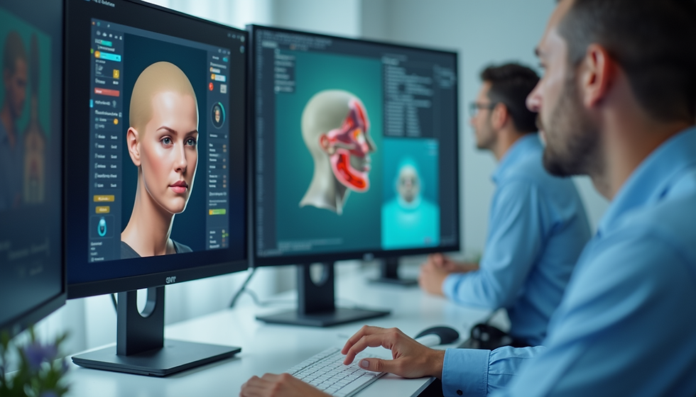
Three-dimensional virtual surgical planning (VSP) allows the surgical team to study the facial skeleton, the teeth and the bite on a digital model before surgery. This article explains how 3D planning works in facial asymmetry, what information it uses, how the plan is transferred to the operating room, and what its limits are.
If you would like to read about the causes, diagnosis and general treatment options first, see our article Facial Asymmetry: Causes, Diagnosis and Surgical Treatment Options.
Why facial asymmetry is a three-dimensional problem
When we look at a face, we usually notice the most visible sign, such as a chin that is off-centre. Underneath, several skeletal differences may be combined:
Midline deviation: the chin and lower dental midline are shifted to one side.
Occlusal cant: the bite plane is tilted, so the teeth and the corners of the mouth are higher on one side.
Rotation (yaw): one half of the jaw is positioned further forward or backward than the other.
Vertical differences: the lower border of the jaw or the jaw angles are at different heights.
Soft tissues can partly hide or exaggerate these skeletal differences. For this reason, correcting only the most visible feature, for example moving the chin alone, may not address the underlying cause. A three-dimensional analysis helps to show which components are present and how much each one contributes.
What information is used for 3D planning
Virtual planning combines several records into one digital model of the patient:
Clinical examination: facial proportions, the facial midline, smile and tooth display, jaw movements and the jaw joints are assessed in person.
Facial photographs: standardised frontal, profile and oblique photographs help to compare the digital plan with the patient's real appearance.
CBCT imaging: cone-beam computed tomography provides a three-dimensional image of the facial bones and teeth.
Digital dental models: an intraoral scan or scanned plaster models give a more detailed picture of the teeth than CBCT alone and are used to plan the final bite.
Bite registration: records the relationship between the upper and lower teeth.
These records are merged so that the bones, teeth and bite can be evaluated together in the correct head orientation.
How virtual surgical planning works step by step
1. Setting the reference position
The digital head model is oriented according to the patient's natural head position and facial midline. This step is important in asymmetry, because measurements made from an incorrect reference can make the face look more or less asymmetric than it really is.
2. Analysing the asymmetry
The surgeon evaluates the position of the upper jaw, lower jaw and chin in all three planes and identifies which components of the asymmetry are skeletal and which are dental.
3. Simulating the surgery
The planned bone cuts are simulated on the computer. Depending on the case, this may involve the upper jaw (Le Fort I osteotomy), the lower jaw (bilateral sagittal split osteotomy, BSSO), both jaws, and/or the chin (genioplasty). The segments are moved virtually to correct the midline, the cant and the rotation while establishing a stable bite.
4. Checking the result
The simulated position is reviewed for facial balance, the bite, contact between the bone segments and, where relevant, changes in the airway. The plan can be adjusted several times before a final decision is made.
5. Transferring the plan to surgery
A digital plan is only useful if it can be reproduced in the operating room. For this purpose, 3D-printed surgical splints are commonly produced from the plan to guide the position of the jaws. In selected cases, cutting guides or patient-specific fixation plates may also be used. The choice depends on the individual case.
What 3D planning can and cannot do
Three-dimensional planning offers clear advantages in asymmetry cases:
It helps to identify all components of the asymmetry, not only the most visible one.
It allows different surgical options to be compared before surgery.
It supports the accurate transfer of the plan to the operating room.
It makes it easier to explain the plan to the patient and the orthodontist.
It also has limits that patients should know about:
No face is perfectly symmetrical, and the aim of treatment is a balanced, harmonious result rather than perfect symmetry.
Soft-tissue changes can be estimated, but the way the lips, cheeks and skin adapt varies from person to person.
A simulation is a planning tool, not a guarantee of the final appearance.
Asymmetry that comes mainly from the soft tissues may require additional or different approaches.
Is 3D planning necessary for every patient?
The level of planning is decided individually. In Dr. Emrah Dilaver's practice, every treatment plan is developed using clinical examination, CBCT imaging and virtual surgical planning according to each patient's needs. In asymmetry cases, three-dimensional analysis is particularly valuable because the problem itself is three-dimensional.
Frequently asked questions
Does 3D planning replace the clinical examination?
No. The digital model is built from records, but decisions about facial balance, smile and tooth display still depend on a careful in-person examination.
Can my orthodontist be involved in the planning?
Yes. In most orthognathic cases, orthodontic preparation and the surgical plan are coordinated, and the digital plan helps both teams work towards the same final bite.
Can I see my plan before surgery?
The planned movements can be shown and discussed during consultation. It is important to remember that the images represent a plan, not an exact prediction of the final appearance.
Can asymmetry be evaluated online before travelling?
A preliminary assessment can be made from photographs, imaging and medical records. Final treatment planning requires an in-person clinical examination.
Next step
If you are concerned about facial asymmetry, you can request a preliminary assessment. Briefly describe your concern in the form; after your request has been reviewed, you can securely share your photographs, imaging and relevant medical records.
This article provides general educational information and does not replace an individual medical assessment.


Comments