
Sleep Apnea Surgery Planning A Surgeon Led Guide to OSA Evaluation

Obstructive sleep apnea is not a jaw problem in every patient. It is a sleep-related breathing disorder that can involve the nose, soft palate, tongue, throat muscles, body weight, sleep position, and facial skeleton. That is why a careful evaluation must come before any discussion of jaw surgery.
From an oral and maxillofacial surgeon’s perspective, the goal is not simply to “make the airway bigger.” The goal is to understand why the airway collapses during sleep, whether jaw position contributes to that collapse, and whether surgery would be appropriate when compared with other treatments.
A responsible approach to sleep apnea surgery planning combines sleep medicine data, symptoms, facial and bite analysis, CBCT imaging, dental scans, airway anatomy, and multidisciplinary judgment. No single scan, number, or measurement can diagnose obstructive sleep apnea or determine the right treatment on its own.
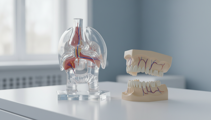
OSA evaluation begins with sleep study findings and symptoms
Obstructive sleep apnea, often called OSA, occurs when the upper airway repeatedly narrows or collapses during sleep. Breathing may pause or become shallow, oxygen levels can drop, and the brain may briefly wake the body to reopen the airway.
The diagnosis is usually made with an objective sleep test, such as an in-lab polysomnography or a home sleep apnea test when appropriate. A surgeon should review this information before considering jaw surgery.
Key findings often include:
Apnea-hypopnea index
This estimates how many breathing interruptions occur per hour of sleep.
Oxygen desaturation
This shows whether oxygen levels drop during sleep and how often.
Arousal pattern
Frequent brief awakenings may fragment sleep, even when the patient does not remember waking.
Sleep position and sleep stage effects
Some patients worsen mainly when sleeping on the back or during REM sleep.
Central versus obstructive events
Jaw surgery is aimed at obstructive collapse. It is not a treatment for central sleep apnea.
Symptoms also matter. Common symptoms include loud snoring, witnessed pauses in breathing, choking or gasping during sleep, morning headaches, dry mouth, unrefreshing sleep, daytime sleepiness, poor concentration, and mood changes. Some patients have significant OSA with few symptoms, while others feel very unwell with milder sleep study numbers.
This is one reason treatment planning must look at the whole person. A sleep report is essential, but it does not explain the full anatomy behind the obstruction.
Jaw position can influence the upper airway
The jaws help support important soft tissues of the upper airway.
The mandible, or lower jaw, carries the tongue through its muscular attachments. When the lower jaw sits far back, the tongue may also sit farther back, especially during sleep when muscle tone decreases. The maxilla, or upper jaw, contributes to nasal and palatal support. The position and width of the upper jaw can influence nasal airflow, dental arch form, and the space behind the soft palate.
This relationship is the anatomical basis for sleep apnea jaw surgery, including maxillomandibular advancement. By advancing the upper and lower jaws in selected patients, the surgeon can move the soft palate, tongue base, and related tissues forward. This may reduce airway collapsibility during sleep.
Yet anatomy is not always simple. A patient may have OSA with normal jaw position. Another patient may have a small or retruded jaw but only mild symptoms. Some patients have obstruction mainly at the soft palate, others at the tongue base, nose, lateral throat walls, or multiple levels.
A surgeon-led assessment asks several questions:
Is the upper jaw retruded, narrow, vertically long, or asymmetric?
Is the lower jaw retruded or small?
Does the bite suggest a skeletal jaw discrepancy?
Is the chin position related to airway support or facial balance?
Are there signs of nasal obstruction or mouth breathing?
Is airway narrowing likely skeletal, soft tissue related, or both?
The answer is rarely based on one finding. It comes from connecting the sleep study with the clinical examination and imaging.

Clinical assessment looks at the face, jaws, bite, and airway
Before imaging or virtual planning, a surgeon examines the patient clinically. This step is central because scans do not replace physical assessment.
A typical oral and maxillofacial evaluation may include:
Facial proportions from the front and side
Chin and jaw projection
Upper and lower jaw relationship
Dental occlusion, meaning how the teeth meet
Overbite, overjet, open bite, crossbite, or crowding
Smile line and incisor display
Temporomandibular joint symptoms
Nasal breathing and septal or turbinate concerns
Mouth opening and tongue size
Tonsil size when visible
Soft palate length and throat appearance
The bite is especially important in surgical planning. Maxillomandibular advancement (MMA) moves the upper and lower jaws, but it must also preserve or correct dental function. If the teeth do not meet properly after surgery, chewing, speech, jaw joint comfort, and long-term stability can be affected.
Facial balance also matters. Jaw advancement for sleep apnea should not be planned as a generic forward movement. The direction and amount of movement may affect lip support, smile aesthetics, chin projection, nasal base shape, and facial harmony.
For international patients, this is why remote review can be useful for initial orientation, but it cannot fully replace a detailed clinical examination when surgery is being considered.
CBCT and 3D airway analysis are useful but limited
Cone beam computed tomography, or CBCT, provides three-dimensional imaging of the facial skeleton and airway space. It can show the jaws, teeth, nasal cavity, sinuses, temporomandibular joints, and upper airway boundaries with useful detail for surgical planning.
In OSA assessment, 3D airway analysis may help the surgeon see:
Narrow areas behind the soft palate or tongue
Jaw relationships in three dimensions
Dental roots and bone anatomy
Skeletal asymmetry
Nasal and maxillary anatomy
Estimated airway volume and minimum cross-sectional area
These details can be very helpful. They allow the surgeon to understand the patient’s anatomy more clearly than with two-dimensional X-rays alone.
Yet CBCT has important limitations.
A CBCT scan is usually taken while the patient is awake and upright or seated. OSA happens during sleep, when muscle tone changes and the airway behaves dynamically. A still scan cannot show how the airway collapses throughout the night.
Airway volume measurements can also vary depending on head posture, tongue position, breathing phase, segmentation method, and software settings. A larger measured volume does not always mean normal sleep breathing. A smaller measured volume does not automatically prove OSA.
CBCT airway measurements alone cannot diagnose obstructive sleep apnea, and they cannot decide treatment by themselves.
The sleep study diagnoses and grades the breathing disorder. CBCT helps explain anatomy and supports planning when surgery is being evaluated.

When maxillomandibular advancement may be considered
Maxillomandibular advancement (MMA) is a jaw surgery that moves both the maxilla and mandible forward. In selected patients with obstructive sleep apnea, this can enlarge and tension the upper airway by advancing the skeletal framework attached to the soft palate, tongue, and throat tissues.
MMA may be considered when:
OSA is objectively confirmed with a sleep study
Symptoms or health risks justify active treatment
Non-surgical treatments are not tolerated, not effective enough, or not suitable
Jaw position appears to contribute to airway narrowing
The patient has a skeletal pattern that may benefit from advancement
The patient understands the risks, recovery, and alternatives
A multidisciplinary team agrees surgery is reasonable
MMA is not the right first step for every patient. Many people with OSA do well with other treatments, such as continuous positive airway pressure, oral appliance therapy, weight management, positional therapy, nasal treatment, or soft tissue procedures when appropriate.
Surgery may be more relevant in patients with moderate to severe OSA, significant jaw retrusion, craniofacial restriction, or persistent OSA despite well-managed non-surgical care. Still, the decision must be individualized. The presence of sleep apnea does not automatically mean the patient needs jaw surgery.
Virtual surgical planning combines anatomy, bite, and airway goals
Modern orthognathic planning uses digital tools to connect multiple sources of information. For OSA patients, this can include CBCT imaging, intraoral dental scans, bite registration, facial photographs, clinical measurements, and sleep study findings.
The surgeon evaluates how the jaws can be moved while respecting:
Dental occlusion
Facial proportions
Chin and lip support
Nasal base changes
Airway anatomy
Jaw joint position
Bone thickness and dental root location
Surgical stability
Patient-specific risks
Virtual surgical planning can simulate jaw movements before surgery. It can help the team plan osteotomy positions, splint design, segment movement, rotation, and fixation strategy. It may also help show how different movement patterns could affect facial balance and airway space.
For example, two patients may both need forward jaw movement, but their plans may be very different.
One patient may need more lower jaw advancement because the mandible is retruded. Another may need counterclockwise rotation of the jaw complex to improve chin projection and airway support. A third may require careful upper jaw movement because of bite, smile, nasal, or facial proportion concerns.
The amount of advancement is not chosen from a standard number. More movement is not always better. Too little may fail to address the anatomical concern. Too much may create facial imbalance, bite problems, lip strain, nasal changes, or instability.
A surgeon-led plan balances airway goals with structure and function. This is where experience in orthognathic surgery, facial analysis, and occlusion becomes essential.
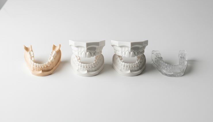
Other treatments and multidisciplinary care remain essential
OSA care should not be isolated within one specialty. A strong treatment plan may involve a sleep physician, oral and maxillofacial surgeon, orthodontist, ENT specialist, dentist trained in sleep medicine, nutrition or weight management support, and primary care physician.
Common non-surgical or less invasive treatments include:
CPAP or other positive airway pressure therapy
This remains a common first-line treatment for many patients.
Mandibular advancement oral appliances
These may help selected patients by holding the lower jaw forward during sleep.
Weight management
In patients where weight contributes to airway collapse, medical weight management can reduce OSA severity.
Positional therapy
Some patients have OSA mainly while sleeping on the back.
Nasal treatment
Treating nasal obstruction may improve comfort with CPAP or oral appliances.
ENT or soft tissue procedures
These may be appropriate when obstruction is mainly related to tonsils, palate, or nasal anatomy.
A surgical consultation should compare these options honestly. Jaw surgery has potential benefits for carefully selected patients, but it also involves anesthesia, swelling, dietary changes, temporary numbness, bite changes, recovery time, and possible complications. Good planning includes both what surgery might improve and what it cannot promise.
Follow-up must include objective sleep reassessment
Postoperative evaluation should not rely only on snoring improvement or how the airway looks on imaging. Many patients feel better after successful treatment, but symptoms alone are not enough to measure OSA control.
Follow-up after jaw surgery may include:
Healing and bite checks
Facial swelling and nerve recovery monitoring
Orthodontic coordination when needed
Nutrition and jaw function guidance
Assessment of nasal breathing and sleep symptoms
Repeat sleep testing after adequate healing
Objective sleep reassessment is important because it confirms how breathing changes during sleep. A repeat sleep study, either in-lab or home-based when appropriate, can measure residual OSA and guide further care.
Some patients may still need CPAP, oral appliance therapy, weight management, or other treatment after surgery. This does not mean surgery has failed in every case. OSA is often multifactorial, and long-term management may require more than one approach.
FAQ
Can a CBCT scan diagnose sleep apnea?
No. CBCT can show airway anatomy, jaw position, and estimated airway dimensions, but it cannot diagnose OSA. Diagnosis requires an appropriate sleep study interpreted in the context of symptoms and medical history.
Does every patient with OSA need jaw surgery?
No. Many patients are treated successfully with CPAP, oral appliances, weight management, positional therapy, nasal treatment, or other approaches. Jaw surgery is considered only when the full evaluation supports it.
What is maxillomandibular advancement?
Maxillomandibular advancement, or MMA, is a surgery that moves the upper and lower jaws forward. In selected OSA patients, it may improve airway support by advancing the tissues attached to the jaws.
How is the amount of jaw advancement chosen?
The movement is individualized. The surgeon considers the sleep study, facial proportions, bite, CBCT findings, dental scans, airway anatomy, jaw joints, and stability. There is no single standard advancement that fits every patient.
Will sleep apnea be cured after jaw surgery?
No treatment can be guaranteed. Some patients have major improvement, while others may have residual OSA and need ongoing care. Objective sleep testing after recovery is needed to assess the result.
Medical disclaimer
This article is for general educational information only and does not replace medical advice, diagnosis, or treatment. Obstructive sleep apnea should be evaluated by qualified healthcare professionals. Any decision about sleep apnea treatment or jaw surgery must be based on an individual assessment, appropriate sleep testing, clinical examination, imaging, and multidisciplinary review.



Comments