
Maxillary Retrusion Explained Recessed Upper Jaw Signs and Treatment Options

A person may look as if the lower jaw is too prominent, when the real issue is that the upper jaw sits too far back. This is one reason maxillary retrusion can be confusing for patients. The face, bite, smile, lips, and nose all influence how the jaws are perceived.
Maxillary retrusion means the upper jaw, also called the maxilla, is underdeveloped or positioned behind the lower jaw. It may be described as a recessed upper jaw or maxillary deficiency. In some cases, the teeth are involved. In others, the main problem is skeletal, meaning it comes from the jaw bones rather than tooth position alone.
This article explains the signs, diagnosis, and treatment options in clear terms. It is for general education only and does not replace a consultation with a qualified orthodontist or oral and maxillofacial surgeon.
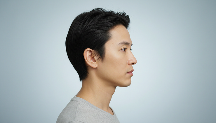
What maxillary retrusion means
The maxilla forms the middle part of the face. It supports the upper teeth, upper lip, cheek area, base of the nose, and part of the eye sockets. When the maxilla is retruded, the midface may look flatter and the upper teeth may sit behind the lower teeth.
Maxillary retrusion can range from mild to severe. Some patients notice only a bite problem. Others notice changes in facial balance, such as reduced cheek support, a flatter upper lip, or a lower jaw that appears strong or protrusive.
Common causes and contributing factors include:
Natural growth patterns
Genetic facial structure
Cleft lip and palate history in some patients
Childhood trauma or surgery affecting jaw growth
Differences in upper and lower jaw development during adolescence
The condition is often discussed as part of a Class III jaw relationship, where the lower teeth or lower jaw sit ahead of the upper teeth or upper jaw.
Common facial and bite features
Maxillary retrusion does not look the same in every patient. The signs can be subtle, especially when the lips, teeth, or lower jaw compensate.
Common facial features may include:
A flatter midface or cheek area
Reduced upper-lip support
A concave facial profile when viewed from the side
A nose that appears more prominent because the upper jaw is set back
A lower jaw that looks strong, long, or forward
Limited upper tooth display when smiling
Deep folds beside the nose and mouth in some cases
Common bite features may include:
Anterior crossbite, where the lower front teeth sit in front of the upper front teeth
Edge-to-edge front teeth
A negative overjet, meaning the upper front teeth are behind the lower front teeth
Posterior crossbite, where upper back teeth sit inside the lower back teeth
Difficulty biting into foods with the front teeth
Uneven tooth wear
Jaw joint or muscle strain in some patients
Some people also report speech or chewing concerns. Others mainly seek evaluation because their facial profile feels unbalanced.
A prominent lower jaw may actually be a recessed upper jaw
Many patients assume that a “big lower jaw” is the cause of a Class III appearance. Sometimes that is true. The mandible, or lower jaw, may be larger or positioned forward.
But in many cases, the lower jaw only looks prominent because the upper jaw is behind it. The face is three-dimensional. If the middle part of the face lacks projection, the chin and lower jaw may appear stronger by comparison.
This distinction matters because treatment planning changes depending on the source of the imbalance.
For example:
Appearance | Possible cause | Treatment direction |
Lower jaw looks prominent | Lower jaw is truly forward | Lower jaw surgery may be considered |
Lower jaw looks prominent | Upper jaw is recessed | Upper jaw advancement may be considered |
Both jaws are imbalanced | Upper jaw is back and lower jaw is forward | Double jaw surgery may be considered |
This is why surgeons do not plan treatment based only on appearance. A full assessment is needed to identify whether the issue is mainly dental, skeletal, or both.

Dental Class III and skeletal Class III are not the same
A Class III problem can be dental, skeletal, or combined.
Dental Class III
A dental Class III problem means the jaw bones may be reasonably balanced, but the teeth are positioned in a way that creates an underbite or crossbite. For example, the upper front teeth may tip backward, or the lower front teeth may tip forward.
In selected dental cases, orthodontic treatment alone may correct the bite. Braces or clear aligners can move teeth into better positions if the bone relationship allows it.
Skeletal Class III
A skeletal Class III problem means the upper and lower jaw bones do not match properly. This may involve a recessed upper jaw, a forward lower jaw, or both.
When the problem is skeletal, orthodontics alone may improve tooth alignment, but it cannot fully reposition the jaw bones. In fact, moving the teeth too far to “hide” the skeletal issue can sometimes create gum, stability, or aesthetic concerns.
Combined Class III
Many patients have both dental and skeletal components. The teeth may have adapted to the jaw imbalance over time. This compensation can make the bite look less severe than the bone problem really is.
Before surgery, orthodontists often “decompensate” the teeth. This means they move the teeth into a healthier position within each jaw, even if the bite temporarily looks worse before surgery. This step helps the surgeon correct the jaw relationship more accurately.
How maxillary retrusion is diagnosed
Diagnosis starts with a clinical examination. The surgeon and orthodontist look at the face, bite, smile, lip posture, airway considerations, jaw movement, and dental health.
A complete assessment may include:
Facial photographs from the front, side, and three-quarter views
Intraoral photographs of the bite
Digital dental scans or impressions
Panoramic X-rays to assess teeth and roots
Cephalometric analysis to measure jaw relationships
CBCT imaging when three-dimensional detail is needed
CBCT, or cone beam computed tomography, gives a 3D view of the jaws, teeth, roots, sinuses, airway, and facial bones. It can be especially helpful when planning orthognathic surgery.
Digital scans show the exact shape and position of the teeth. When combined with CBCT data, they help the team study how the upper and lower jaws fit together.
Modern planning often uses 3D virtual surgical planning. This allows the surgeon to simulate jaw movements before surgery, evaluate symmetry, and plan splints or guides when needed. It also helps the patient understand the proposed changes more clearly.
Can orthodontics alone correct a recessed upper jaw?
Orthodontics can move teeth. It cannot move the adult upper jaw forward in a meaningful skeletal way.
For growing children, certain orthopedic appliances may influence upper jaw growth in carefully selected cases. Timing is important, and results vary depending on growth pattern and severity.
For adults, braces or aligners may help if the problem is mainly dental. If the upper jaw itself is recessed, orthodontics alone can camouflage the bite but cannot correct the bone position.
Camouflage treatment may be suitable for mild cases when facial balance is acceptable and the bite can be made stable. It may not be ideal when there is:
A significant underbite
A concave facial profile
Poor upper-lip support
Limited upper tooth display
Severe dental compensation
A bite that cannot be corrected without unhealthy tooth movement
The decision should be made after combined orthodontic and surgical assessment.

When Le Fort I maxillary advancement may be considered
A Le Fort I osteotomy is the standard surgical technique used to reposition the upper jaw. During this procedure, the surgeon carefully separates the maxilla, moves it into the planned position, and fixes it with small plates and screws.
When the upper jaw is moved forward, the procedure is called maxillary advancement. This may be considered when the main issue is maxillary deficiency and the lower jaw is reasonably positioned.
Le Fort I maxillary advancement may help correct:
Skeletal underbite caused by a recessed maxilla
Poor upper tooth display caused by upper jaw position
Reduced midface and upper-lip support
Certain crossbites or open bites, depending on the case
Facial imbalance related to the upper jaw
The movement is planned in millimeters, but the effect is broader than a number. A small movement can affect the bite, smile, lip support, nasal base, and profile. This is why planning must be detailed and individualized.
When double jaw surgery may be necessary
Some patients need surgery on both jaws. This is called double jaw surgery, or bimaxillary orthognathic surgery.
Double jaw surgery may be considered when:
The upper jaw is recessed and the lower jaw is also forward
There is significant facial asymmetry
The bite cannot be corrected predictably with one jaw movement
The smile line, chin position, or facial proportions require changes in both jaws
Airway and functional factors support a two-jaw plan
For example, if the surgeon advances only the upper jaw in a patient whose lower jaw is also too far forward, the bite or profile may still not be balanced. In that case, moving both jaws can create a better relationship between the teeth, lips, chin, nose, and face.
Double jaw surgery is more complex than single-jaw surgery, but it may give better control in selected cases. The goal is not to make every face look the same. The goal is to create a stable bite and natural facial balance.
How upper jaw advancement can affect the face
Upper jaw surgery changes more than the bite. Because the maxilla supports the middle third of the face, advancement can influence several visible areas.
Facial profile
Advancing the upper jaw can reduce a concave profile. The midface may look more supported, and the lower jaw may appear less dominant even if the lower jaw is not moved.
Smile
Maxillary advancement can improve the relationship between the upper teeth and lower teeth. In some cases, it can also improve upper tooth display when smiling. If vertical changes are needed, the surgeon may adjust how much the upper jaw moves up or down as part of the plan.
Upper-lip support
A recessed maxilla can leave the upper lip looking flat or unsupported. Moving the upper jaw forward may improve lip projection and soft tissue support.
Nose and nasal base
Because the upper jaw forms part of the base of the nose, maxillary advancement can affect nasal shape slightly. The nasal base may widen, and the nasal tip or upper lip angle may change. Surgeons use specific techniques to help control these changes, but the exact soft tissue response varies from person to person.
Patients should discuss these aesthetic effects before surgery. A good surgical plan looks at function and appearance together.

General recovery after upper jaw surgery
Recovery varies by patient, procedure, and whether one or both jaws are treated. The first days usually involve swelling, congestion, limited mouth opening, and a soft or liquid diet.
General recovery often includes:
A short hospital stay or monitored recovery period, depending on the clinic and case
Swelling that increases during the first few days, then gradually improves
Nasal stuffiness after upper jaw surgery
Temporary numbness in the upper lip, cheeks, teeth, or gums
A soft diet while the bone heals
Elastic bands to guide the bite
Follow-up visits with the surgical and orthodontic team
Gradual return to normal activity based on the surgeon’s instructions
Most patients need orthodontic treatment before and after surgery. Braces or aligners help prepare the teeth before surgery and refine the bite afterward.
Numbness often improves over time, but nerve recovery can be slow and is not always complete. Infection, bleeding, relapse, sinus issues, and bite changes are possible risks. A surgeon should explain the risks, expected recovery, and alternatives before treatment.
Short FAQ about maxillary retrusion
Is maxillary retrusion the same as an underbite?
Not always. Maxillary retrusion means the upper jaw is set back. An underbite describes how the teeth meet. A recessed upper jaw can cause an underbite, but an underbite can also come from lower jaw position or tooth position.
Can braces fix a recessed upper jaw?
Braces can move teeth, but they cannot significantly move the adult upper jaw forward. If the problem is skeletal, surgery may be needed to correct the jaw position. Mild dental cases may be treated with orthodontics alone.
Is Le Fort I osteotomy painful?
Patients are under anesthesia during surgery and do not feel the procedure. After surgery, discomfort, swelling, pressure, and congestion are common. Pain is usually managed with medication and careful follow-up.
Will upper jaw surgery change my nose?
It can. The upper jaw supports the base of the nose, so advancement may slightly widen the nasal base or change the nasal tip and upper lip angle. Surgeons plan for this and may use techniques to help control nasal changes.
Do all Class III patients need double jaw surgery?
No. Some need only upper jaw surgery, some need lower jaw surgery, and some need both. The decision depends on the position of each jaw, the bite, the facial profile, symmetry, airway factors, and treatment goals.
Key takeaway
Maxillary retrusion can affect the bite, smile, lips, nose, and overall facial balance. A lower jaw that looks too prominent may actually be the result of an upper jaw that sits too far back. The most reliable way to understand the problem is through a full clinical examination, dental analysis, CBCT imaging when appropriate, and digital treatment planning.
Orthodontics alone may help in mild dental cases. Skeletal cases often require orthognathic surgery, such as Le Fort I maxillary advancement, and some patients may need double jaw surgery for the most balanced correction.



Comments