
Sleep Apnea Jaw Surgery Guide: MMA Surgery, Airway Surgery, and 3D Planning

Obstructive sleep apnea can look like “just snoring” from the outside, but the real problem happens inside the airway. During sleep, breathing repeatedly slows or stops because soft tissue and anatomy narrow the airway. Over time, this can affect energy, concentration, blood pressure, heart health, metabolic health, and overall quality of life.
For some people, the main issue is not only weight, nasal blockage, or soft tissue collapse. The position of the upper jaw, lower jaw, chin, tongue, and facial skeleton can leave too little room behind the tongue and soft palate. In selected cases, sleep apnea jaw surgery may help by moving the jaw framework forward and increasing airway space.
This guide explains how obstructive sleep apnea is evaluated, when jaw anatomy matters, what maxillomandibular advancement involves, and how 3D planning supports safer, more personalized treatment. It is informational only and does not replace a consultation with a qualified sleep medicine physician, oral and maxillofacial surgeon, orthodontist, or ENT specialist.

What obstructive sleep apnea means for health
Obstructive sleep apnea, often called OSA, occurs when the upper airway becomes partly or fully blocked during sleep. The brain senses reduced airflow or lower oxygen levels and briefly wakes the body enough to reopen the airway. These events can happen many times per hour, even if the person does not remember waking up.
Common signs may include:
Loud snoring
Pauses in breathing witnessed by a partner
Gasping or choking during sleep
Morning headaches
Dry mouth on waking
Daytime sleepiness
Poor focus or memory
Irritability or low mood
High blood pressure that is hard to control
OSA severity is usually measured through a sleep study. One key number is the apnea-hypopnea index, or AHI. It estimates how many breathing interruptions occur per hour of sleep.
Typical AHI ranges are:
AHI range | General category |
Fewer than 5 events per hour | Usually considered within normal range |
5 to 14.9 events per hour | Mild OSA |
15 to 29.9 events per hour | Moderate OSA |
30 or more events per hour | Severe OSA |
AHI is useful, but it is not the whole story. Oxygen levels, sleep quality, symptoms, medical conditions, anatomy, and treatment tolerance all matter when building a treatment plan.
How jaw position and facial anatomy affect the airway
The airway is not a rigid pipe. It is a soft, flexible space surrounded by muscles, the tongue, the palate, the jaws, and the neck. During sleep, muscles relax. If the airway is already narrow, this relaxation can allow the tongue or soft palate to fall backward and block airflow.
The lower jaw and tongue are closely linked
The tongue attaches to the lower jaw and nearby structures. When the lower jaw sits far back, the tongue base can also sit farther back. This can reduce the space behind the tongue, especially when lying flat during sleep.
A small or retruded lower jaw may contribute to:
Narrowing behind the tongue
Mouth breathing
Posterior tongue collapse during sleep
Difficulty using some oral appliances
A facial profile with a recessed chin or weak lower jaw support
This does not mean every person with a small jaw has sleep apnea. It means jaw position can be one part of a larger airway picture.
The upper jaw can affect nasal and palatal space
The upper jaw, or maxilla, supports the nasal floor, upper teeth, cheeks, and part of the palate. A retruded or narrow upper jaw may affect nasal airflow and the space behind the soft palate.
In some patients, both jaws sit behind an ideal functional position. When the upper and lower jaws are moved forward together, the soft tissues attached to them can also shift forward. This is the basic idea behind maxillomandibular advancement.

When jaw anatomy can lead to airway obstruction
Jaw anatomy is more likely to play a role when the facial skeleton limits the airway space even before sleep begins. During sleep, muscle relaxation can turn that limited space into repeated obstruction.
Clinical findings that may raise suspicion include:
A small lower jaw or recessed chin
A retruded upper jaw
A long lower facial height with clockwise jaw rotation
A narrow palate or dental arch crowding
A high tongue position relative to the airway
A bite problem linked to skeletal imbalance
Persistent OSA despite other treatments
Difficulty tolerating CPAP or oral appliance therapy
Some people seek jaw surgery for sleep apnea after years of symptoms and failed treatment attempts. Others are diagnosed during evaluation for jaw imbalance, bite problems, or facial asymmetry. The key is to determine whether the airway issue truly relates to skeletal anatomy.
How sleep apnea is diagnosed and evaluated
A careful evaluation comes before any discussion of obstructive sleep apnea surgery. Surgery should not be based on snoring alone or on facial appearance alone.
Sleep studies and AHI
A sleep study records breathing patterns, oxygen levels, heart rate, sleep stages, body position, and breathing events. It may be done in a sleep lab or, for selected patients, with a home sleep apnea test.
A sleep report usually includes:
AHI
Oxygen desaturation levels
Time spent below normal oxygen saturation
Sleep position data
Snoring data
Central apnea events, if present
Sleep efficiency and arousals
This information helps the team understand the type and severity of sleep apnea. It also helps distinguish obstructive sleep apnea from central sleep apnea, which has a different cause and treatment approach.
Clinical examination
A clinical evaluation may include the nose, mouth, throat, bite, jaw joints, facial profile, dental arches, and neck. Specialists may assess tonsil size, soft palate shape, tongue volume, nasal septum position, turbinate size, and jaw relationship.
Many patients benefit from a multidisciplinary review. This may involve sleep medicine, oral and maxillofacial surgery, orthodontics, ENT, pulmonology, and anesthesia.
What maxillomandibular advancement surgery does
Maxillomandibular advancement, often called maxillomandibular advancement or MMA surgery, is a form of airway surgery that moves both the upper jaw and lower jaw forward. The goal is to enlarge and stabilize the upper airway by advancing the skeletal framework and the soft tissues attached to it.
In broad terms, MMA may:
Increase space behind the soft palate
Increase space behind the tongue
Reduce airway collapsibility during sleep
Improve facial skeletal support in selected patients
Address certain bite and jaw relationship problems at the same time
Surgeons usually perform MMA through incisions inside the mouth, so visible facial scars are often avoided. The bones are repositioned and secured with small plates and screws. The exact movements depend on the patient’s anatomy, bite, airway findings, facial balance, and treatment goals.
MMA is not a quick or minor procedure. It requires careful planning, anesthesia assessment, recovery time, and follow-up sleep evaluation. For the right candidate, it can be an important sleep apnea treatment option, especially when non-surgical care is not enough or cannot be tolerated.
How MMA differs from conventional orthognathic surgery
MMA and conventional orthognathic surgery can use similar surgical techniques. Both may involve upper jaw surgery, lower jaw surgery, or chin-related procedures. The difference is the main purpose and planning focus.
Conventional orthognathic surgery | MMA for sleep apnea |
Often focuses on bite correction, facial balance, or jaw deformity | Focuses on improving airway space while also respecting bite and facial balance |
Dental occlusion is usually the central planning guide | Airway volume, airway shape, sleep study results, occlusion, and facial aesthetics all guide planning |
Advancement may be limited by facial and bite goals | Advancement is often planned with airway improvement as a major goal |
Sleep testing may not be part of the workup unless symptoms suggest OSA | Sleep testing and airway analysis are central parts of the evaluation |
A patient may need both functional bite correction and airway improvement. In these cases, the surgical plan must balance occlusion, temporomandibular joint health, facial proportions, nasal breathing, and airway goals.
Why CBCT and 3D planning matter
Modern evaluation often includes cone beam computed tomography, known as CBCT. This scan creates a 3D view of the facial bones, teeth, nasal airway, and upper airway space.
CBCT does not replace a sleep study, because sleep apnea happens during sleep and involves dynamic soft tissue collapse. Still, CBCT can show airway anatomy in ways that regular 2D X-rays cannot.
3D airway analysis
A 3D airway analysis can help the team examine airway volume, narrow points, skeletal relationships, jaw position, and dental alignment. It can also support better communication between the surgeon, orthodontist, and patient.
The scan may help identify:
A narrow airway behind the soft palate
A narrow airway behind the tongue
Upper jaw retrusion
Lower jaw retrusion
Dental arch limitations
Facial asymmetry
Chin and jaw rotation patterns
These findings must be interpreted with the clinical exam and sleep study. A narrow airway on imaging does not automatically mean surgery is required.
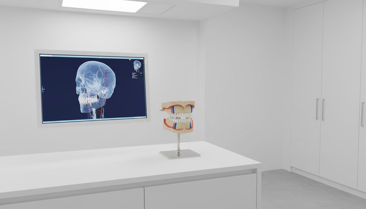
3D virtual surgical planning
With 3D virtual surgical planning, the surgical team can simulate jaw movements before surgery. This helps plan how far and in what direction the jaws may move.
Planning may include:
Upper jaw advancement
Lower jaw advancement
Counterclockwise rotation of the jaw complex
Chin position changes, if needed
Dental midline correction
Bite setup
Custom guides or splints for surgery
How surgeons balance airway, bite, and facial appearance
Moving the jaws forward can affect the bite and the face. A good plan does not treat the airway as separate from the rest of the facial skeleton.
Dental occlusion guides the final fit
Dental occlusion means how the upper and lower teeth meet. If the jaws are advanced without respecting occlusion, the patient may end up with chewing problems, bite instability, or jaw joint strain.
Some patients need orthodontic treatment before MMA. Braces or aligners may prepare the teeth so they fit correctly after jaw movement. In other cases, surgery-first planning may be possible, but this depends on the bite and the team’s protocol.
Facial balance still matters
Airway improvement is a key goal, but the face must remain balanced. The surgeon considers the nose, lips, chin, cheek support, smile display, and facial proportions. The goal is not simply to move the jaws as far forward as possible. The goal is an individualized movement that supports breathing, function, and a natural facial balance.
This is especially relevant for international patients considering sleep apnea surgery in Istanbul or any other destination. Travel plans should never replace a complete workup, clear communication, and structured follow-up care.
Who may be a candidate for sleep apnea jaw surgery
Jaw surgery is usually considered when OSA is moderate to severe, when anatomy supports a skeletal cause, or when other therapies are not effective or not tolerated. It may also be considered in selected mild cases with significant symptoms and clear jaw-related obstruction, but this requires careful review.
Possible candidates include people with:
Diagnosed obstructive sleep apnea
Jaw retrusion or skeletal narrowing related to airway obstruction
Poor tolerance of CPAP despite support and adjustment
Limited benefit from oral appliance therapy
OSA combined with a significant bite or jaw relationship problem
A desire for a more anatomy-based option after full counseling
Not all patients need surgery. Some have OSA mainly related to weight, nasal obstruction, tonsil enlargement, alcohol or sedative use, sleep position, or soft tissue collapse without major skeletal restriction. Others do well with non-surgical treatment and should continue it.
Good candidate selection is one of the most important parts of jaw surgery for sleep apnea.
Non-surgical and less invasive treatment options
Many patients start with non-surgical care. These treatments may be effective, especially when the OSA is mild to moderate or when the anatomy does not point to a major skeletal cause.
Common options include:
CPAP therapy CPAP uses gentle air pressure to keep the airway open during sleep. It is a standard treatment for many patients with OSA.
Oral appliance therapy A custom mandibular advancement device can hold the lower jaw forward during sleep. It may help selected patients, especially those with mild to moderate OSA.
Weight management when relevant Weight is not the only cause of OSA, but it can affect airway narrowing in some people.
Positional therapy Some patients have more events while sleeping on the back. Positional treatment may reduce events in selected cases.
ENT treatment Nasal surgery, tonsil surgery, or soft palate procedures may help when the blockage is mainly in those areas.
Lifestyle changes Reducing alcohol before bed, reviewing sedating medications with a physician, and improving sleep habits may support treatment.
A complete plan may combine therapies. For example, nasal treatment may improve CPAP comfort, or orthodontics may prepare a patient for MMA surgery.
What to expect before surgery and during recovery
Before surgery, the care team usually confirms the diagnosis, reviews medical history, studies imaging, evaluates the bite, and discusses risks and benefits. Patients may need dental records, facial photographs, CBCT imaging, sleep study reports, blood tests, anesthesia clearance, and orthodontic planning.
The surgical plan should explain:
Which jaws will be moved
Approximate direction and size of movement
Expected bite changes
Whether orthodontics is needed
Possible effects on facial appearance
Recovery timeline
Risks, limits, and alternatives
After surgery, swelling, congestion, facial tightness, temporary numbness, and a modified diet are common. Many patients start with liquids or soft foods and slowly return to normal chewing as healing allows. Follow-up visits track bone healing, bite stability, oral hygiene, nerve sensation, and airway symptoms.
Recovery varies. Some people return to light daily activities within a few weeks, while full healing and bite settling take longer. Air travel, strenuous activity, and return to work should be discussed with the surgical team.

Follow-up sleep testing is part of the treatment
Symptom improvement can be encouraging, but follow-up testing gives objective information. A repeat sleep study may be recommended after healing to measure AHI, oxygen levels, and sleep quality after surgery.
Follow-up also helps identify whether more treatment is needed. Some patients may still need CPAP at a lower pressure, an oral appliance, nasal care, weight-related support, or positional therapy. Others may need long-term monitoring if symptoms return or health conditions change.
The best care is individualized. OSA is a medical condition with many possible causes, and no single treatment fits every patient.
A multidisciplinary plan gives the clearest path forward
Sleep apnea treatment works best when the team looks at the whole person, not just one scan or one symptom. Sleep physicians measure the breathing problem. Surgeons evaluate skeletal anatomy. Orthodontists protect the bite. ENT specialists assess the nose and throat. Anesthesia teams review surgical safety.
For selected patients, obstructive sleep apnea surgery with maxillomandibular advancement can address the skeletal framework that contributes to airway collapse. For others, CPAP, oral appliances, ENT care, lifestyle support, or combined treatment may be the better route.
The real next step is a careful diagnosis. A sleep study, airway-focused exam, CBCT assessment, and thoughtful discussion of all options can show whether MMA belongs in the treatment plan, or whether a less invasive path makes more sense.



Comments